
What is Spinal Stenosis?
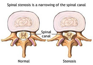
Spinal stenosis is a narrowing of the open spaces within your spine, which can put pressure on your spinal cord and the nerves that travel through the spine to your arms and legs. It occurs most often in the lower back and the neck.
While spinal stenosis may cause no signs or symptoms in some people, other people may experience pain, tingling, numbness, muscle weakness, and problems with normal bladder or bowel function.
Spinal stenosis is most commonly caused by wear-and-tear changes in the spine related to osteoarthritis. In severe cases, doctors may recommend surgery to create additional space for the spinal cord or nerves.
Symptoms of Spinal Stenosis
Many people have evidence of spinal stenosis on X-rays, but may not have signs or symptoms. When symptoms do occur, they often start gradually and worsen over time. Symptoms vary, depending on the location of the stenosis:
- In the neck (cervical spine). Cervical stenosis can cause numbness, weakness or tingling in a leg, foot, arm or hand. Tingling in the hand is the most common symptom, and many people also report problems with walking and balance. Nerves to the bladder or bowel may be affected, leading to incontinence.
- In the lower back (lumbar spine). Compressed nerves in your lumbar spine can cause pain or cramping in your legs when you stand for long periods of time or when you walk. The discomfort usually eases when you bend forward or sit down.
When to see a doctor
Make an appointment with your doctor if you have persistent pain, numbness or weakness in your back, legs or arms.
Causes of Spinal Stenosis
While some people are born with a small spinal canal, most spinal stenosis occurs when something happens to reduce the amount of space available within the spine. Causes of spinal stenosis may include:
- Overgrowth of bone. Wear and tear damage from osteoarthritis on your spinal bones can prompt the formation of bone spurs, which can grow into the spinal canal. Paget’s disease, a bone disease that usually affects adults, also can cause bone overgrowth in the spine.
- Herniated disks. The soft cushions that act as shock absorbers between your vertebrae tend to dry out with age. Cracks in a disk’s exterior may allow some of the soft inner material to escape and press on the spinal cord or nerves.
- Thickened ligaments. The tough cords that help hold the bones of your spine together can become stiff and thickened over time. These thickened ligaments can bulge into the spinal canal.
- Tumors. Abnormal growths can form inside the spinal cord, within the membranes that cover the spinal cord or in the space between the spinal cord and vertebrae.
- Spinal injuries. Car accidents and other major trauma can cause dislocations or fractures of one or more vertebrae. Displaced bone from a spinal fracture may damage the contents of the spinal canal. Swelling of adjacent tissue immediately following back surgery also can put pressure on the spinal cord or nerves.
Most people with spinal stenosis have passed the age of 50. When younger people develop spinal stenosis, the cause is typically a genetic disease affecting bone and muscle development throughout the body.
Complications with Spinal Stenosis
Rarely, untreated cases of severe spinal stenosis may progress and cause permanent:
- Numbness
- Weakness
- Balance problems
- Incontinence
- Paralysis
Diagnosing Spinal Stenosis
Spinal stenosis can be difficult to diagnose because its signs and symptoms resemble those of many age-related conditions. Imaging tests may be needed to help pinpoint the true cause of your signs and symptoms.
Imaging tests
These tests may include:
- X-rays. Using a small exposure to radiation, X-rays can reveal bony changes, such as bone spurs that may be narrowing the space within the spinal canal.
- Magnetic resonance imaging (MRI). In most cases, this is the imaging test of choice for diagnosing spinal stenosis. Instead of X-rays, an MRI uses a powerful magnet and radio waves to produce cross-sectional images of your spine. The test can detect damage to your disks and ligaments, as well as the presence of tumors. Most important, it can show pressure on the spinal cord or spinal nerves.
- CT myelogram. If you can’t have an MRI, your doctor may recommend computerized tomography (CT), a test that combines X-ray images taken from many different angles to produce detailed, cross-sectional images of your body. In a CT myelogram, the CT scan is conducted after a contrast dye is injected. The dye outlines the spinal cord and nerves, and it can reveal herniated disks, bone spurs and tumors.